


RETINA SERVICES IN KAKINADA
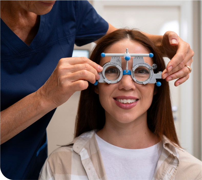
If you are looking for a retina hospital in Kakinada, you are at the right place. We offer specialized retina eye care to address various retinal conditions. Our experienced retina specialists are dedicated to preserving and restoring vision, providing comprehensive treatments for age-related retinal issues, diabetes, hypertension, and more. Trust our experts for your retina health needs.
Retina is a thin nerve tissue that lines the back of the eye. It is responsible for the sense of light and creates impulses that travel to our brain through the optic nerve. It is equivalent to the sensor of a camera. It is the most important part of the eye and is responsible for vision. Hence any damage to the retina can result in permanent damage to the eye and result in total loss of vision.
Age, diabetes, hypertension, myopia and trauma are a few conditions that can predispose an eye to retinal disease. It is very important to visit a retina specialist immediately if you notice sudden loss of vision, reddish hue in vision, sudden appearance of dark spots or threads or insect like objects moving in your visual field or wavy or distorted vision to name a few.
INVESTIGATIONS
FUNDUS FLUORESCEIN ANGIOGRAPHY (FFA)
This investigative procedure comprises of injecting a dye called fluorescein into one of the veins in your arm and taking rapid serial photographs of its passage within the delicate blood vessels of the eye in the retina and choroid, using a digital Fundus camera.
The information obtained from this test aids your doctor in making a diagnosis and planning treatment like laser or intravitreal injections.
Apart from the needle prick and the light flash of the camera there is no discomfort from this test. A few patients may have nausea and by remaining calm and taking deep breaths help overcome it. Your usual diet can be taken soon after the procedure. Fluorescein is a non-toxic drug. In rare instances, it can produce an allergic reaction, which responds rapidly to appropriate medication. Serious life-threatening allergic reactions, though exceptionally rare, can occur. The skin and urine may appear yellow in colour for about 36 hours following the test and is not a cause of any concern.
You must be accompanied by an adult attendant during this test.
OPTICAL COHERENCE TOMOGRAPHY (OCT)
It is a non-invasive, non-contact imaging technology, with ultra-high speed 27,000 axial scans per second. It provides high-resolution 5-µm axial and 15-µm transverse resolution in tissue.
The procedure requires you to sit in front of the OCT machine and place your chin on chin rest and forehead touching the head support. You are required to focus at a given target and keep your eye still while the scan is being performed.
The layers within the retina can be differentiated and the retinal thickness can be measured. Also, the change from the previous visit, the effect of treatment can be analyzed.
B-SCAN
B SCAN is a two-dimensional imaging system which utilizes high-frequency sound waves ranging from 8-10 MHz. It is used for imaging of intraocular structures and giving information about the status of the lens, vitreous, retina, choroid, and sclera.
B-scan ultrasound is most useful when direct visualization of intraocular structures is difficult or impossible in cases like corneal opacities, miosis, pupillary membranes, dense cataracts, or vitreous opacities.
In many instances, ultrasound is used for diagnostic purposes even though pathology is clinically visible. Such instances include differentiating intraocular tumors, rhegmatogenous versus exudative retinal detachments, and disc drusen versus papilledema.
RETINAL TREATMENT PROCEDURES
Intravitreal Anti-VEGF injections:Intravitreal Anti-VEGF injections are the first line of therapy in a variety of retinal conditions like diabetic macular edema, cystoid macular edema in vascular occlusions, wet age related macular degeneration, polypoidal choroidal vasculopathy etc.
Intravitreal steroid injections:They are used to treat uveitic cystoid macular edema, recurrent or macular edema not responding to anti-VEGF injections.
RETINAL LASERS
Pan retinal photocoagulation: This usually involves multiple sessions spaced 10-15 days apart and are used to treat conditions like proliferative diabetic retinopathy, vein occlusions etc.
Focal laser: This is usually a single session and a short procedure used to treat conditions like central serous retinopathy, diabetic macular edema etc
RETINAL SURGERIES:
Retinal surgeries are carried out to treat certain conditions like retinal detachments, epiretinal membranes, macular holes, vitreous hemorrhage and others.
Book an Appointment
Have any questions or feedback, Feel free to reach out to us. We always available to help