


CORNEA CLINIC
The cornea is the clear front surface of the eye and it allows light to enter the eye. Diseases that affect cornea are second only to cataract in major causes of blindness all over the world. Corneal diseases like corneal abrasions, pterygium, dry eye, keratitis and keratoconus require specialist management. If left untreated they can result in compromised vision. A few symptoms caused by corneal diseases are pain, foreign body sensation, blurred vision, redness, watering and sensitivity to light.
Our corneal surgeon is highly experienced in treating various corneal conditions that include anterior segment trauma, ocular surface diseases including dry eye, contact lens related eye conditions, microbial keratitis management and refractive surgery.

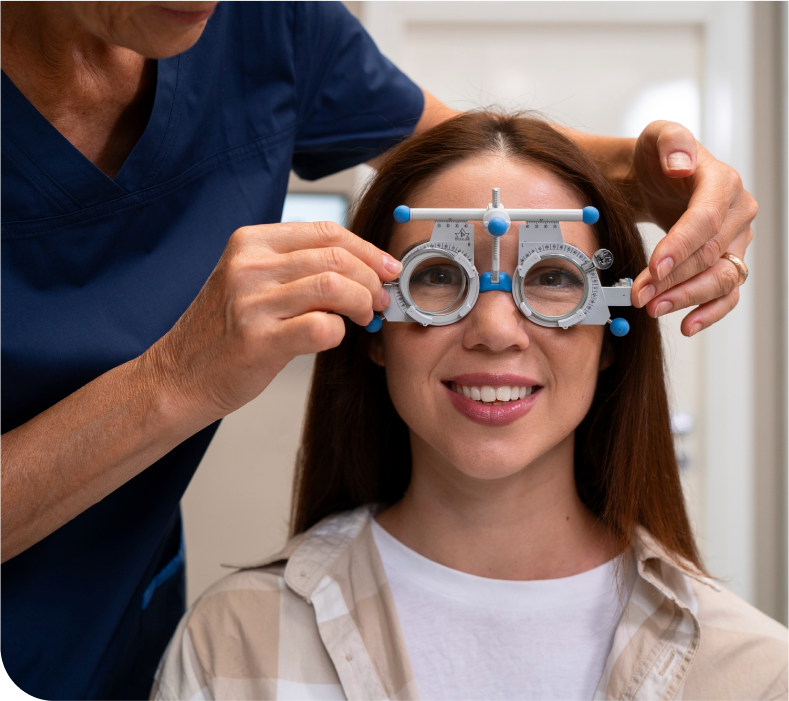
INVESTIGATIONS
CORNEAL TOPOGRAPHY (PENTACAM)
Corneal topography is a technology that creates 3D maps of the surface curvature of the cornea. Corneal topography produces a detailed, visual description of the shape and power of the cornea. This analysis provides your doctor with very fine details of the cornea used to diagnose, monitor, and treat various eye conditions. They are also used in fitting contact lenses and for planning surgery, including laser vision correction like PRK and LASIK.
SPECULAR MICROSCOPY
It is a technology used to assess the morphology, density and function of corneal endothelial cells. Endothelium is the innermost of the 5 layers of cornea.
Common ocular conditions, such as glaucoma, uveitis and Fuchs endothelial dystrophy, may produce changes in the structure and function of the corneal endothelium that result in corneal edema and visual impairment. Additionally, clinical circumstances, such as contact lens wear and intraocular surgery, may compromise the endothelium and cause corneal edema. An accurate diagnosis of endothelial disease may be the key in not only determining cause of corneal edema, but also developing a treatment plan.
CORNEAL SURGICAL PROCEDURES
PTERYGIUM SURGERY:
Pterygium surgery is a procedure performed to remove the conjunctival growth on the cornea. A pterygium is a growth of scar-like tissue of the conjunctiva onto the edge of the cornea. It typically occurs on the nose side of the eye but can also occur on the ear side. Small pterygia are common and often don’t cause significant symptoms. They can cause redness and irritation. If they get large, they can also affect the vision. If they cause decreased vision or severe discomfort, they can be surgically removed.
The pterygium is excised and some conjunctival tissue from the upper part of the eye is removed and placed in the area from which the pterygium was removed, to decrease the chance of recurrence of the pterygium. However, the pterygium can occasionally recur. Pterygium surgery is performed using local or topical anesthesia.
CORNEAL COLLAGEN CROSS LINKING:
Corneal collagen cross-linking or C3R is a treatment for an eye problem called Keratoconus In this condition, the front part of your eye, called the cornea, thins out and gets weaker over time. This makes it bulge into a cone shape, which can distort your vision and make it hard to see. If the symptoms of keratoconus get severe, you will need a corneal transplant. In corneal cross-linking, doctors use eyedrop medication and ultraviolet (UV) light from a special machine to make the tissues in your cornea stronger. The goal is to keep the cornea from bulging more. It’s called “cross-linking” because it adds bonds between the collagen fibers in your eye. They work like support beams to help the cornea stay stable. Corneal cross-linking is the only treatment that can stop progressive keratoconus from getting worse. And it may help you avoid a corneal transplant, which is major surgery.
Book an Appointment
Have any questions or feedback, Feel free to reach out to us. We always available to help